By Dr. Barry Hartup, ICF Director of Veterinary Services
An old English proverb states “the eyes are the window to the soul.” In my line of work, the eyes are sometimes a window into the health of a patient. Though the eyes may not be the most prominent feature of cranes, compared to raptors for instance, the cranes do show beautiful diversity in eye color, and have a predictable shape and relationship to the form of the skull. When changes occur in this presentation, the observant among us know something could be wrong.
Such was the case on February 18th when ICF aviculturist Kyle Tainter called late in the afternoon to say that Omega, ICF’s exhibit male whooping crane, suddenly had reddish looking eyes, quite different than the normal pale yellow color. A diagnosis of hyphema, or blood in the front chamber of the eyes, was made. Omega’s eyes were also more rounded in shape and projecting more from the skull than normal. Omega was subdued and apparently blind or nearly so. He was locked into a warmed shelter with his mate, Seurat, in order to prevent further injury, and monitored closely.
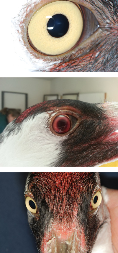
Photos, right, from top to bottom: A normal Whooping Crane eye, from the female Seurat; Omega’s right eye following injury. There is a red haze of blood in front of the eye and the pupil is dilated. The left eye was similarly affected; Omega two weeks after his injury. His right eye is still a bit misshapen and dilated than his left, which now appears normal. Photos by Dr. Ellison Bentley and Kyle Tainter
This condition is often associated with a traumatic blow, but could also be due to more insidious causes such as underlying infection or poisoning. Thankfully for Omega, there was no evidence of underlying disease or likelihood of exposure to any kind of anticoagulant substance. Unfortunately, there is no specific course of treatment for hyphema; helping the patient with the pain and discomfort from the increased pressure in the eye (think glaucoma) and resulting inflammation is the first order of business. His attitude improved over the course of days with pain relief and anti-inflammatory medication, and by two weeks, we allowed the pair access back into their outdoor winter quarters. Omega’s visual capacity had improved, but some minor changes in eye shape and pupil response to bright light were still present. A full ophthalmic examination by University of Wisconsin School of Veterinary Medicine colleagues Drs. Ellison Bentley and Gillian McLellan revealed evidence of small hemorrhages in the retina of the right eye but normal intraocular pressure, meaning no lasting glaucoma.
We believe Omega should make a near full recovery with no significant loss of vision — he is seeing small food items and beating his mate to the smelt we provide in preparation for the breeding season. Come pay him a visit beginning in April. He’d love to see (!) you.
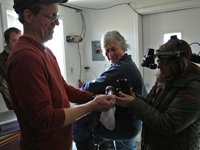
Dr. Gillian McLellan (right) obtains a pressure reading on Omega’s left eye, while aviculturist Marianne Wellington and Dr. Hartup restrain him for examination. Aviculturist Kyle Tainter, who deftly discovered Omega’s problem, watches. Photo by Dr. Ellison Bentley